

+91 - 9818704499
+91 - 9311444806
+91 - 9311444806
Care Well Medical Centre is coming up with latest art of treatment in management of stable vitiligo. We are routinely doing Melanocyte transplantation for leucoderma patients . This treatment is done on day care procedure and does not require admission. In this treatment a thin skin graft is taken and treated chemically to extract melanin which is pigment responsible for skin coloration.
The melanin is separated from the solution and transplanted to the area where there is loss of pigment. The depigmented area takes up melanin and its discoloration improves over 3 to 6 months. Previously we used to practice skin grafting on this area white patches. The disadvantage with skin grafting was a donor area of equivalent size was required. This means for a 100 square centimeter of white patch we needed 100 square centimeter of skin graft. In melanocyte transplant we need only 10 to 20 square centimeter of donor area. In skin grafting the colour match is not always optimal and the marks of graft are always left. In melanocyte transplant the cosmetic results are superior and at times the colour match is gratifying. Often it becomes difficult to point out if any one has undergone this procedure.
Normally upto 100 square centimeter of white patch can undergo melanocyte transplantation. In our Care Well Medical Centre we have covered more than 300 square centimeter area in single sitting.
The white patches (vitiligo ) of finger tips, toes etc is called Acral Vitiligo This is most difficult to treat. We are using Blister grafting and punch grafting in the acral vitiligo. So combination of Melanocyte transplant and blister grafting we can manage any vitilgo which is medically stable.
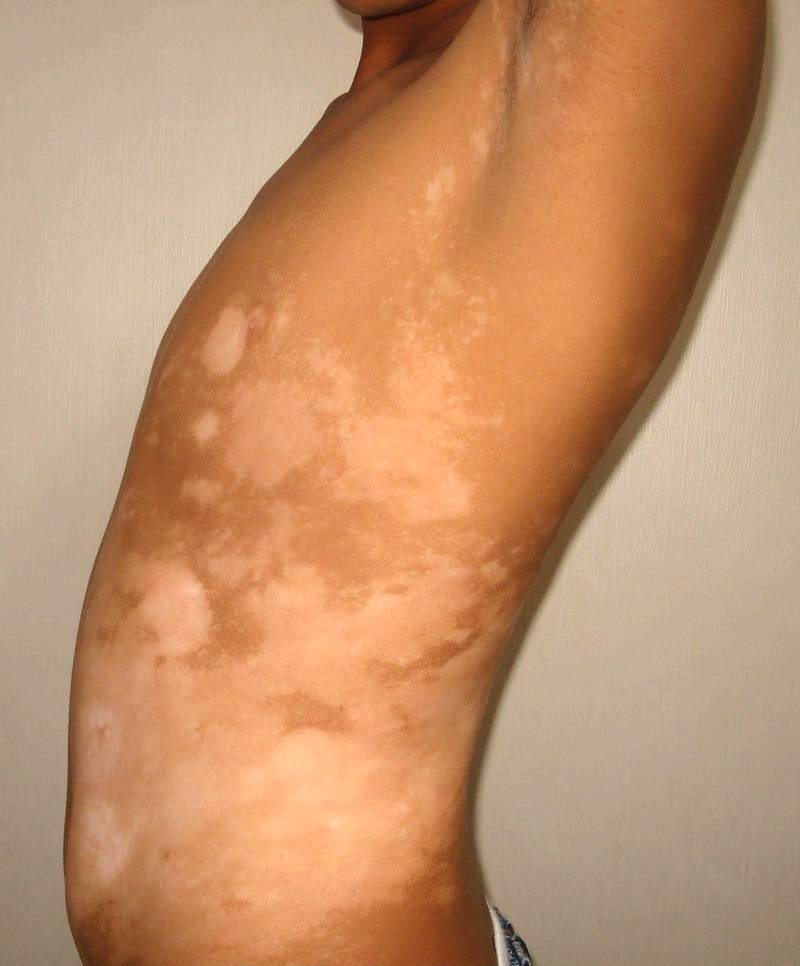
Vitiligo is a pigmentation disorder in which melanocytes in the skin - tissues that line the inside of the mouth, nose, the genital areas, the rectal areas (mucous membranes) and the retina of the eyes - are destroyed as a result of which white patches of skin appear on different parts of the body. The term Vitiligo is probably derived from the latin word Vitilus - meaning calf and was first used by roman physician Celsus of 1st century AD,The characteristics white patches of disease resembled the white patches of a spotted calf in India. Leukoderma is a generic name for relatively or absolutely lightened in colour.
Symptoms of Vitiligo The disease is characterised by the appearance of depigmented patches(milky white) on the skin, common in sun exposed areas like hands, feet, arms, face and lips. Other common areas include armpits, groin, around the mouth, eyes, nostrils, navel and genitals. Rarely the patches show slight erythema, but as a rule they show only depigmentation and sensivity to light, the hair may be white or black but in a particular lesion, when hairy areas are involved the hair may turn white.
Focal Pattern :Depigmentation is limited to one or only few areas.
Segmental Pattern : Depigmentation develops on only one side of the body.
Generalised Pattern : Depigmentation develops on different parts of the body.
The primary goal is to restore the function of the skin & improve the patient's appearance. The choice of therapy depends upon the number of patches, the size of patch & on patient's preference for the treatment.
» Topical steriod therapy.
» Topical psoralen photochemotherapy.
» Oral psoralen photochemotherapy.
» Oral steroid therapy.
» Depigmentation.
» Autologous skin Grafts.
» Punch Grafts.
» Autologous Melanocyte transplantation.
Care Well Medical Centre melanocyte transplantation requires no hospitalisation & is done on an out patient basis at his surgical center. It requires only local anesthesia and patient is sent home immediately after the procedure. After anaesthetising the normal skin (usually from thigh/buttocks) a 4cm by 2cm skin is removed, using Silver skin grafting knife which gives excellent thin grafts containing only the epidermis. This skin is then incubated in trypsin for one hour & at the end of one hour, dermis if found is separated from epidermis. The epidermis is then divided into small pieces and mixed with minimum essential media, which keeps the cells alive till it is transplanted onto the body, centrifuged repeatedly till a melanocyte pellet is obtained. From a 4x2cm piece of skin, upto 100square-centimeters of depigmented patches can be treated. The depigmented patch is anaesthetized (Xylocaine 2% or Emla ointment) & dermabrasion is done using either a laser or micromotor to remove epidermis, taking care not to injure the dermis, which can result in scarring. Once the epidermis has been removed, the melanocyte suspension is applied over the patch & wound is covered with a Collagen dressing. This dressing is then removed after 7 days
» Treated areas remain pink after removal of dressing.
» Earliest pigmentation is seen within 3-6 weeks.
» Entire area covers up in 3 to 4 months.
» Excellent cosmetic results are seen in 5-6 months.
» There were no cosmetic side-effects after the procedure.some patients may require more than one sitting for 100% pigmentation.
» Day care procedure (no admission is required irrespective of site of patch)
» Very large areas can be treated.
» Pigmentation appears rapidly within 3-6 weeks.
» Cosmetic results are excellent.
» Difficult areas like fingers, lips can also be treated(some patients may require two sittings for complete pigmentation)
» No scarring seen after surgery.
 Increasing number of men and women who want to get rid of droopy upper eyelids and lower lid bags are achieving rejuvenated and young they desire from a special cosmetic surgery
 We, a team from Care well Medical Centre have been in Hair Transplantation for last 10 years. Initially FUT or Strip method was used. From last 4 years FUE is gaining popularity.
 Care Well Medical Centre is a renowned cosmetic surgery, plastic surgery and hair treatment center in South Delhi in CR Park touching nearby areas
 Carboxy therapy mentioned to the administration of carbon dioxide gas in cutaneous and subcutaneous nerves for therapeutic purposes.
 A fat grafting process involves transfers of fat from excess fat areas i.e. outer thighs in and injects it into areas that may be lacking in volume i.e. face, hands, breasts or buttocks.
 Cavitation is body sculpting with out anesthesia, with out scars, with out discomfort, with out down-time and presents a risk-free substitute to liposuction.
 CARE WELL MEDICAl CENTRE conducts Camp in Mandakani Welfare association, Chitranjan Park on 16th August 2014. The camp was headed by Dr. Sandeep Bhasin and his team.
 Even now, after all these years, she can realize the moment when she first spotted a numb skin patch on her back.
 Address: Care Well Medical Centre, 1, NRI Complex, Chittaranjan Park, New Delhi - 110019
Phone: +91-9818704499, +91-9311444806
Email: info@carewellmedicalcentre.in
Copyright © 2014, Carewell Medical Centre. All Rights Reserved

